Birds’ respiratory systems differ from that of mammals, such as the unidirectional airflow that occurs through the lungs. The avian respiratory system is used for gaseous exchange, vocalization, and thermal regulation.1 It includes a complex but efficient respiratory cycle which, along with other unique aspects of avian respiration, is the topic of this article.
Nostrils & Nasal Cavity
The birds’ nostrils or ‘nares’ (i.e., nasal openings) and the adjacent regions inside the nasal cavity vary among the different species.1 Often, the nares of birds’ are located at the base of the beak in a dorsal, ventral, or lateral position but are situated at the tip of the beak in kiwis, flightless birds endemic to New Zealand.1 Additionally, some birds, such as the seabirds, gannets, and boobies, have no external nares but breathe through a narrow gap between the distal upper and lower beak, enabling them to dive at high speeds without water blowing out of their nostrils.2 In general, the avian nasal cavity is compressed laterally and divided medially by a very thin septum. This cartilaginous structure separates the right and left sides of the nasal cavity. Nostrils that are separated from each other by a septum are therefore imperforate.2 However, the nares of some birds, such as vultures, are not divided by a septum. In these birds, the nares are joined, and the nares of vultures are described as perforate nostrils because a septum does not separate them. In some species like psittacines (e.g., parrots), the nares have a thick cere, a waxy fleshy covering. Many birds also have a hard, keratinized structure called the operculum at the center of each nare. This structure acts as a baffle to prevent inhalation of foreign bodies.2
Glottis & Trachea
Birds can breathe through their nose or mouth.2 Air enters through the nares into the conchae, complex structures forming the upper chamber of the nasal cavities in nearly all birds. Birds have three conchae (e.g., rostral, middle, and caudal) that act as intermittent countercurrent heat exchangers during routine lung ventilation.3,4 The nasal passages and sinus communicate with the choana, an anatomic cleft located in the roof of the mouth.4 During respiration, air moves across the pharynx, the part between the mouth and esophagus, to the glottis, opening to the trachea. The glottis abuts the choana and is located at the base of the tongue.4 It is usually accessible in awake birds, and the proximal trachea attaches to the glottis. The trachea is formed from a series of complete tracheal cartilage that forms complete cartilaginous rings, in which rigid interlocking rings cannot expand.5 In addition, birds need long necks to help manipulate objects with their beaks, and in some species, the trachea is so long that it forms coils (e.g., the trumpeter swan).2 Birds have a trachea of wider diameter than mammals of equivalent size, resulting in a dead space, the inhaled volume of air that does not take part in gas exchange. The dead space in birds is 4.5 times that of mammals of equivalent size. Birds have an increased tidal volume, the amount of air that moves in or out of the lungs with each respiratory cycle.2 Tidal volume in birds provides air equivalent to a complete turnover of the airspace of the lungs plus trachea. Birds have a slower and deeper rate of breathing than mammals. For example, birds like swans breathe slowly, 10 breaths per minute, whereas the ostrich has a slower breathing rate at 3 to 5 breaths per minute.2 This contrasts with normal dogs and cats that generally have a breathing rate of between 15-30 breaths per minute while resting or sleeping.
The Syrinx
The syrinx, from the Greek word for panpipe, occurs just cranial to the heart at the junction located where the trachea bifurcates (or splits). 2, 4 It can be classified as tracheal, tracheobronchial, or bronchial based on location.2 Most birds are described as having a tracheobronchial syrinx since it is based on both tracheal and bronchial elements.1 The syrinx is analogous to a mammalian larynx in which sounds are produced but differs in that the syrinx lacks vocal cords that vibrate with air currents and is rudimentary in species like vultures and ostriches.4 It consists of modified tracheobronchial cartilages, two vibrating tympaniform membranes, and muscles that vary the membrane tension.2 The thin tympaniform membranes line the medial and lateral bronchi, large air passages that lead from the trachea to the lungs. Sound is produced during expiration by the vibration of air through the syrinx.2 The number of syringeal muscles varies with species, and muscles are used to cause vibration to produce sound. 1, 2, 4 For example, parrots have three pairs, hawks have only one pair, while the songbird has five pairs.1, 2 Importantly the syrinx is a common site of obstruction by foreign bodies like seeds or fungal granulomas.2, 4 Because of this, loss of voice, change in voice, or dyspnea, difficult or labored breathing should always indicate to the owner to seek veterinary care that includes evaluation of the syrinx.5
Lungs & Air Flow Through the Respiratory Tract
The respiratory system in birds plays an essential role in thermoregulation, maintaining normal body temperature.6 Additionally, it delivers oxygen from the air to the tissues and removes carbon dioxide.2,6 Birds have a much higher oxygen demand than mammals due to flight and the ability to fly at altitude. The lungs of birds are 10 times more efficient than mammal lungs in capturing oxygen.2, 6 The avian lungs are dorsal in position, attached to the thoracic ribs and vertebra, filling the intercostal spaces, the anatomic space between two ribs.1, 5 The lungs of birds are small and thus undergo a slight change in volume during breathing. They are more rigid than mammalian lungs because they contain more cartilage, making the lungs of birds less compressible.1 As previously mentioned, the trachea of birds narrows and bifurcates where the syrinx is the narrowest portion of the trachea.4 The tracheal bifurcation creates the beginning of the left and right primary bronchi, the main airways that branch off into smaller and smaller passageways. All bronchi are lined by smooth muscle, causing dilation and contraction, and each primary bronchus enters one lung.2, 4 After entering the lungs, each bronchus divides, giving rise to four sets of secondary bronchi according to the lung area they supply (e.g., mediodorsal, medioventral, laterodorsal, and lateroventral).2 The secondary bronchi further subdivide into the tertiary bronchi called parabronchi. Most of the parabronchi are a parallel series of hundreds of narrow tubes. The inner surfaces of parabronchi are pierced by numerous openings into individual chambers called atria from which air capillaries extend.2, 6 Inspired air moves from the parabronchi into the atria which open along the walls of the parabronchi before flowing into air capillaries.4 The air capillaries are the avian analog of alveoli, tiny air sacs in the lungs allowing rapid gaseous exchange. Unlike mammalian alveoli, however, air capillaries communicate with each other. Air capillaries are where gas exchange occurs within the parabronchi and surrounding mantle of vascular-rich tissue.6 There are two types of parabronchial tissue in the avian lung called paleopulmonic parabronchial tissue and neopulmonic parabronchial tissue.2, 7, 6 The paleopulmonic and neopulmonic parabronchi are histologically indistinguishable from each other, but the gas flow direction differs within the two types of the lung.2, 6 Paleopulmonic parabronchial tissue is found in all birds. It consists of parallel, minimally anastomosing parabronchi or paleopulmonic bronchi that make up the bulk of lung tissue.2, 6 Neopulmonic parabronchial tissue is a meshwork of anastomosing parabronchi that are irregularly branched. They do not comprise more than 25% of the parabronchi and are located in the caudolateral portion of each lung.2, 6.
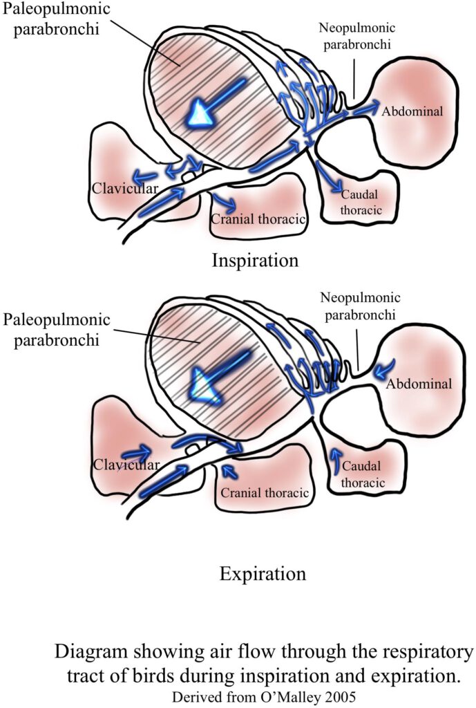
Air Sacs
The avian respiratory system includes a series of air sacs that constitute 80% of the respiratory volume and extends from the body cavity into the wing, vertebra, and leg bones. 2, 5 They are thin, distendable, transparent sacs that are lined by simple squamous epithelium with few blood vessels.2, 8 The air sacs store and pump air through the stationary lungs and are not involved in oxygen exchange. Instead, air sacs act as bellows to direct airflow into and through the lungs in only one direction, which is essential for maximizing oxygen extraction.6,4, 2 This process allows birds to take in oxygen even during exhalation and breathe at much higher elevations than mammals because of their more efficient lung structure.6 In addition, air sacs help to reduce the amount of heat produced during flight by evaporation and play a role in sound production, courtship displays, and some species cooling of the testes for spermatogenesis.2 In most birds, there are three pairs of air sacs and two single ones creating eight in total, but in some species, the cervical sacs are paired, making nine air sacs.2 The lungs directly connect to the air sacs, of which there are 4-5 sets (e.g., cervical, interclavicular, cranial thoracic, caudal thoracic, and abdominal).4 Notably, the cervicocephalic air sac communicates with the infraorbital sinus and not directly with the lungs.4, 5 In an area called the ostium, air sacs connect to the secondary bronchi in which the parabronchi interconnect with all the air sacs except the cervical one.2 It is thought that the shape and alignment of the secondary bronchi create a pressure differential between the cranial and caudle air sacs influencing airflow through the parabronchi.2


The Mechanics of Respiration
Birds do not possess a muscular diaphragm that separates the abdominal and thoracic cavity.5, 6 Instead, they have the horizontal septum that separates the lungs from the viscera and plays no active role in respiration but passively helps displace the viscera during breathing.2
Birds have the same abdominal muscles as mammals (although they are smaller because the sternum is so prominent) and depend on cervical, thoracic and abdominal muscles for inspiration and expiration.4, 6 Both inspiration and expiration require active muscle contraction.2 Importantly, because breathing is achieved by muscles that move the ribs and sternum, it is crucial that birds not be restrained across the chest.5 During inspiration, the inspiratory muscles contract. In this process, the internal volume of the thoracoabdominal cavity increases along with the volume of the air sacs, which are the only significant volume-compliant structures within the body cavity.6 Air flows from the atmosphere into the pulmonary system as the pressure within the air sacs becomes negative relative to ambient atmospheric pressure.1, 6 In this process, the air sacs in birds have a bellows-like action in moving the air between the lungs and air sacs.1 The respiratory cycle of birds’ is complex yet an efficient process where fresh air is ensured to move to the lungs both on inspiration and expiration. 4, 5. Unlike mammals, where ventilation takes one respiratory cycle, it takes two ventilation cycles to move air through the respiratory system of birds.2, 5 In this process, most of the inspired air bypasses the lungs and goes into the caudal thoracic and abdominal air sacs during the first inspiration.4 With the second inspiration, air moves from the lungs into the cranial air sacs. During the first exhalation, air moves from the caudal thoracic and abdominal air sacs into the lungs, where gas exchange occurs. During the second exhalation, air leaves through the trachea.4 Needless to say, the mechanics of avian respiration is complex..
References
- King, A. S., & Jonh Mclelland. (1984). Birds : their structure and function. Baillière Tindall.Millis, D. L., & Levine, D. (2014). Canine rehabilitation and physical therapy (2nd ed.). Saunders, Cop
- Bairbre O’malley. (2005). Clinical anatomy and physiology of exotic species : structure and function of mammals, birds, reptiles, and amphibians. Elsevier Saunders.
- Geist, N. R. (2000). Nasal Respiratory Turbinate Function in Birds. Physiological and Biochemical Zoology, 73(5), 581–589. https://doi.org/10.1086/317750
- Avian respiratory and thoracic surgery (Proceedings). (n.d.). DVM 360. https://www.dvm360.com/view/avian-respiratory-and-thoracic-surgery-proceedings-0
- Scott, D. E., & C.A.B. International. (2021). Raptor medicine, surgery, and rehabilitation. Cabi.
- Inhaled Anesthesia for Birds | IVIS. (2001, March 2). Www.ivis.org. https://www.ivis.org/library/recent-advances-veterinary-anesthesia-and-analgesia-companion-animals/inhaled-anesthesia
- Welty, J.C. and L. Baptista. 1988. The life of birds, fourth edition. Saunders College Publishing, New York, NY.
- Causey, G. (2000). Sturkie’s avian physiology ; 5th ed. Academic.
Figure references
Bairbre O’malley. (2005). Clinical anatomy and physiology of exotic species : structure and function of mammals, birds, reptiles, and amphibians. Elsevier Saunders.
Powell, F.L. 2000. Respiration. Pp. 233-264 in Avian physiology, fifth edition (G. Causey Whittow, ed.). Academic Press, New York, NY.